SiHKAL: Shulgins I Have Known and Loved

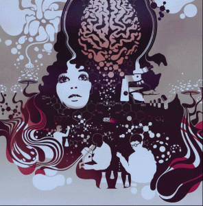
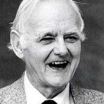
Hamilton Morris, Vice magazine’s emissary for all things mindbending, conducts a new interview with Sasha and Ann Shulgin.


After spending days, weeks, months poring over the work of psychonaut-in-chief, Alexander Shulgin, Hamilton Morris mustered up the chutzpah to give him a call and request an interview. The result is this: an epic love-fest on the man who birthed Ecstasy in a test-tube. Hamilton visits the Shulgin residence (in San Francisco, naturally) and tempers his fanboy freakout with a rare and intensive look at the home and laboratory that caused the balls of millions to trip.

-VBS.TV: SiHKAL: Shulgins I Have Known and Loved

Norepinephrine

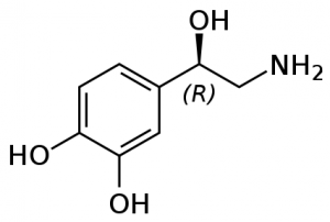
Norepinephrine (NE) is a neurotransmitter, involved in behaviours including alertness, anxiety, and attention. Most norepinephrine cells cluster in a pair of nuclei called the locus coeruleus, Latin for a “blue spot” in the brain stem. Each locus coeruleus is a long tube shaped cluster of norepinephrine neurons, both consisting of only 45,000 to 60,000 nerve cells in total. A second group of nerve cells arises deeper down in the medulla, and delivers norepinephrine mostly to the hypothalamus.

Primate brains have a dense network supplying norepinephrine to the posterior parietal lobe (which integrates sensory information), to part of the thalamus, and to the outer layers of the optic lobes in the midbrain. Relatively little norepinephrine is supplied to the temporal cortex, involved in speech, high-level visual processing (ie facial recognition), and memory. This outlines a distinctive feature of norepinephrine, that it is generally involved in direct sensory input rather than sensory processing. But the hypothalamus (which regulates hunger, thirst, fatigue, and sleep cycles) has the highest concentration of norepinephrine at 1150 nanograms per gram, indicating the vital role this neurotransmitter plays in deep primal instincts.

NE cells in the locus coeruleus fire especially in response to stressful, painful, noxious stimuli. In rats, they fire faster for up to thirty seconds if the toe is compressed in a painful manner. A strong but generic stimuli for cats (say a loud noise) will prompt a brief discharge of norepinephrine, but this response dwindles as the stimuli is repeated and the cat becomes conditioned.

In higher primates like monkeys, pain and stress also cause norepinephrine cells to fire faster. But what really gets monkey norepinephrine production going is fruit juice, their favorite treat. Norepinephrine cells fire at their highest rates when drinking fruit juice, seven to fifteen times per second. The norepinephrine cells of primate brains appear to be slightly different than that of lower animals, as they are especially activated by noxious stimuli and also certain specific attractive stimuli. Norepinephrine cells fire much slower if primates are given opiate based painkillers however, and during dream states (REM) of sleep.

So what exactly happens when norepinephrine cells fire more frequently? They appear to have rather few direct effects, but act primarily as a modulator. For instance, norepinephrine alone does little to change the slow background firing of cells in the hippocampus proper (involved in memory and spatial navigation) - but if these cells have been excited by addition of glutamate, norepinephrine makes them fire even faster. Norepinephrine also makes hippocampal cells fire faster if they have been excited by natural outside visual and auditory stimuli. It also reduces spontaneous background activity, increasing the signal to noise ratio of sensation from outside. This increased signal to noise ratio has become very valuable in treating symptoms of conditions such as ADHD. Norepinephrine and dopamine both play a large role in attention and focus. Various amphetamines and other compounds like methylphenidate (Ritalin) prescribed for ADHD cause higher levels of norepinephrine and dopamine.

Other compounds can act as agonists at norepinephrine receptors. Clonidine is used to relieve the suffering of addicts withdrawing from opiates. It activates certain autoreceptors which slow the norepinephrine cells’ firing rate, reducing symptoms such as anxiety, restlessness, and aching in muscles and joints. This clinical success emphasizes the theories linking excessive norepinephrine activity within the locus coeruleus with states of anxiety and tension. For instance, investigation into patients suffering panic attacks suggests that they may lack the normal mechanisms that would hold their norepinephrine systems in check.

The Neurobiology of Psychedelic Drugs

An excellent review paper has appeared in Nature Reviews: Neuroscience entitled “The neurobiology of psychedelic drugs: implications for the treatment of mood disorders”. Here’s the abstract:

After a pause of nearly 40 years in research into the effects of psychedelic drugs, recent advances in our understanding of the neurobiology of psychedelics, such as lysergic acid diethylamide (LSD), psilocybin and ketamine have led to renewed interest in the clinical potential of psychedelics in the treatment of various psychiatric disorders. Recent behavioural and neuroimaging data show that psychedelics modulate neural circuits that have been implicated in mood and affective disorders, and can reduce the clinical symptoms of these disorders. These findings raise the possibility that research into psychedelics might identify novel therapeutic mechanisms and approaches that are based on glutamate-driven neuroplasticity.

As a result, Nature has decided to highlight some related reading in their Blog Focus: Hallucinogenic drugs in Modern Medicine and Mental Health with four great articles.

The secret history of psychedelic psychiatry Highlighting the early advances of Dr. Humphrey Osmond and other pioneers of psychedelic psychotherapy.
Serotonin, Psychedelics and Depression
Investigates the relationship between the action of psychedelics and current theories of depression, both which rely on the neurotransmitter serotonin.
Ketamine for Depression: Yea or Neigh?
Investigating the potential of the NMDA antagonist ketamine as a breakthrough treatment for depression.
Visions of a psychedelic future
A Western scientist’s experience with the psychedelic brew ayahuasca.

Smoked cannabis for chronic neuropathic pain: a randomized controlled trial

A great paper just came out in the Canadian Medical Association Journal, dealing with the ability of cannabis to combat neuropathic pain. Neuropathic pain is thought to be caused by damage to the nervous system, and often does not respond to conventional painkillers. It is estimated that 1-2% of the adult population is affected, and patients with this chronic pain have reported using smoked cannabis to relieve pain, improve sleep and improve mood.

Suffice to say that most of the time this isn’t suggested by doctors and dispensed by prescription. So does it actually work? 21 adults with post-traumatic or postsurgical neuropathic pain completed the study, and were randomly assigned to receive cannabis at four potencies (0%, 2.5%, 6% and 9.4% THC). They assigned the various strengths of cannabis as part of a “crossover trial”, where each subject would receive one of the concentrations randomly for a two week period. After four two-week periods and random “crossovers” to other potencies, everyone had tried every potency.

Participants inhaled a single 25-mg dose of cannabis through a pipe three times daily for the first five days in each two-week period, followed by a nine-day washout period before the next trial. Daily average pain intensity was measured using an 11-point scale, and effects on mood, sleep, quality of life, and adverse events were recorded.

And the results? Turns out that you need decent pot for pain relief, but it doesn’t have to be an insanely potent strain. Lower potency cannabis did not significantly reduce pain, but the 9.4% THC cannabis reduced pain, improved ability to fall asleep, and improved quality of sleep. The beautiful outcome is that cannabis containing 10% THC is widely available and only 75mg a day (slightly over two grams a month) is needed to cause statistically significant improvements for those suffering chronic pain.

Mark A. Ware, Tongtong Wang, Stan Shapiro, Ann Robinson, Thierry Ducruet, Thao Huynh, Ann Gamsa, Gary J. Bennett, Jean-Paul Collet, Smoked cannabis for chronic neuropathic pain: a randomized controlled trial, Canadian Medical Association Journal, Volume 182, Number 14, 2010, Pages 694-701.

Dopamine

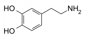
Dopamine (DA) is a neurotransmitter present in many animals, from the honeybee to the dolphin. It is involved in many behaviors associated with motivation and reward. The majority of dopamine in humans is released from two areas.

First is the the substantia nigra, whose darkly pigmented cell bodies supply dopamine to the deep motoric nuclei of the basal ganglia. Dopamine concentrations in one nuclei, the putamen, exceed anywhere else in the brain reaching a level of 5740 nanograms per gram. Dopamine released in this nigrostriatal pathway helps us quickly execute our motor patterns and associated movements. For instance, if dopamine drops to very low levels on one side of this system Parkinsonian symptoms will arise, with sluggish arm and leg movements on one side of the body.

Second is the ventral tegmental system. Its cells send much of their dopamine up to the nucleus accumbens in the ventral striatum, and a separate pathway supplies the limbic system. Two other branches supply dopamine to the cortex in the prefrontal and cingulate regions. An especially dense network of dopamine fibers covers the inner part of the prefrontal lobe, the same region where the thalamus sends its major input. The intersection of these two pathways in the prefrontal cortex remains one of the brain’s most constant features, dating back to the tree shrew and reminding us of our origins in the forest.

Suppose you take a rat, normally a social animal, and reduce its social stimuli over a prolonged period making it a hermit in a cage. The different dopamine systems react in different ways - the metabolic activity of the dopamine cells supplying the frontal lobes slows, whereas metabolism increases in dopamine cells projecting up to the dorsal and ventral striatum. Even though the isolated rats have now quieted down and show few spontaneous behaviors (frontal lobes), they jump more when an electrical shock is delivered to the foot (dorsal and ventral striatum). Increased social exposure causes a reversal of these effects - more active and less nervous rats.

Each person’s brain is unique, as the number and distribution of dopamine receptors varies widely. Early in life, brain development responds to male or female hormones present. Male rats already show more evidence of dopamine receptors in their cortex and amygdala only a few days after being born.

There are five known dopamine subreceptors in humans. The most abundant dopamine subreceptor in the nervous system is the D1 receptor, which regulate neuronal growth and development and mediates some behavioral responses. It also stimulates adenylyl cyclase and activates cyclic AMP-dependent protein kinases, involved in the regulation of glycogen, sugar, and lipid metabolism. D2 receptors are gaining prominence as potential keys to certain diseases, as increased D2 receptor levels have been linked to schizophrenia, certain Parkinson’s symptoms, and narcolepsy.

The major functional role of dopamine systems in the human brain appears twofold. First, there are prominent motoric effects. Decades of research on rats and mice have shown that any cause of indirect release of dopamine into the nucleus accumbens causes rats to engage in specific motor sequences: they move around more, they sniff more, and engage in repetitive grooming behaviors. This is not unlike a human who has experienced dopamine increase caused by cocaine or other drug consumption, as they fidget, sniff and lick their lips, run their fingers through their hair, or pick at the skin.

But there is more than just a general mobilizing or energizing effect. This became clear when experiments were undertaken as animals pressed on a lever, working to receive food as a reward. Drugs which increased dopamine levels caused the animals to become more selective and efficient, suggesting that dopamine helps to sustain goal directed behavior. While dopamine was initially dubbed the “pleasure” neurotransmitter, recent research suggests that pleasure is a less appropriate term than “motivation”. As any speed freak will tell you, after a certain point self-administration of the drug becomes distinctly dysphoric, but the desire to continue dosing remains strong and is not correlated with the level of
“pleasure” (if any) that the next dose will produce.

There are also interesting correlations to personality. “Extroverted” personalities tend to show higher spinal fluid levels of dopamine breakdown products, and a set of aggressive patients also showed the same evidence of higher dopamine turnover. After practicing yoga meditation for six months however, the levels of dopamine breakdown products of the aggressive patients fell from an average of 51 nanograms per liter to 41 nanograms per liter.

The Three Biogenic Amine Systems

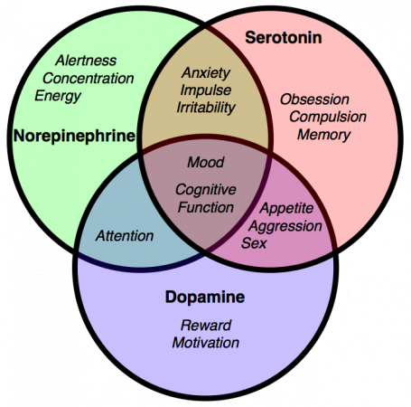
The three potent neurotransmitters dopamine, norepinephrine, and serotonin were known to science prior to the 1960s, but the precise structure and extent of their nerve cells was unclear. Annica Dahlström and Kjell Fuxe used the novel technique of histoflourescence to map the pathways that released them into the brain in a landmark paper in 1964, “Evidence for the existence of monoamine neurons in the central nervous system“. The field exploded, and strange facts emerged. They are greatly outnumbered, as our brain only holds a million or so of them. But they exert a huge influence over the remaining billions of other nerve cells, fanning out into up to 500,000 nerve endings connecting to hundreds of other distant cells.

GABA

Nerve cells are designed to fire repetitively, and are subject to a barrage of stimuli. The brain prevents itself from spiraling out of control into hyperexcitable states by inhibition, a way of “turning down the volume” in the brain. The major workhorse for this is gamma animobutyric acid, or GABA. In a strange parallel, the brain synthesizes GABA in one step from glutamate, the brain’s major excitatory neurotransmitter. Excitation can therefore be efficiently followed by the capacity for inhibition, two naturally opposing processes depending on, and counterbalancing, the other. GABA and glutamate are the yin and yang of our nervous system.

There is hardly a single behavior that does not involve GABA. It is estimated to be involved in one third of all transmissions at synapses, and GABA nerve cells alone make up 40 percent of the total population of nerve cells, with glutamate cells adding perhaps another 30 percent. Broadly, the brain applies its fast acting GABA circuits at all levels to hold in check the local excitatory processes. Some GABA cells inhibit pivotal nerve cells that directly command other essential nerve cell populations. GABA mechanisms hold aggressive behavior in check in many animal species. If rats are made hyperactive by injection of dopamine into the nucleus accumbens, they will quickly quiet down after GABA is injected into the same nucleus.

GABA agonists include alcohol and benzodiazepines such as Xanax. Widely used for their tranquilizing, anti-anxiety, or sleep producing properties, they can create an incredibly risky state of addiction if used too frequently. Unlike other drugs that when withdrawn simply cause the perception of extreme suffering, cold turkey withdrawal causes GABA action to drop precipitously, inducing states of extreme agitation or convulsions, possibly causing death. A slow tapering of the drug, allowing natural GABA to rebound slowly, is the typically recommended course of action.

The inhibitory effects of GABA can be related to the “tunnel vision” experienced during states of drunkenness, where the visual field is reduced and attention is paid only to the objects directly in front of the subject. In contrast, bicluculline is a drug that stops the inhibitory role of GABA. When applied to the visual cortex, it expands the apparent visual fields by three to five times.

A traveller taking a benzodiazepine for a long haul flight and a frat boy on his 21st birthday both may experience periods of retrograde amnesia. This emphasizes the fact that when GABA systems become overactive, they can block the way a person accesses recently produced memories.

The anti-anxiety properties of GABA agonism are not limited to the “higher” civilized frustrations of the overworked executive as well. They can also relieve the deep primal anxiety of a baby monkey separated from his mother. Diazepam, a benzodiazepine, quickly stops their agitated behavior and cries of distress. High brain levels of dopamine are reduced, as well as high blood levels of ACh and cortisol which are correlated with stressful experiences.

Acetylcholine

Acetylcholine (ACh) was the first neurotransmitter to be discovered. Identified in the year 1914 by Henry Hallett Dale for its actions on heart tissue, it was confirmed as a neurotransmitter by Otto Loewi. Both received the 1936 Nobel Prize in Medicine for their work. The nerve cells that release ACh are widespread and influential, involved in alertness, arousal, memory, and the fast brain waves which occur during waking and REM sleep.

In the peripheral nervous system, acetylcholine activates muscles, and is a major neurotransmitter in the autonomic nervous system. In the central nervous system, acetylcholine and the associated neurons form a neurotransmitter system, the cholinergic system, which is associated with anti-excitatory actions. ACh is also involved with synaptic plasticity, specifically in learning and short-term memory.

There are two main types of ACh receptors.

Nicotinic Acetylcholine Receptors

The first type of ACh receptor is called nicotinic, from the familiar compound nicotine of the tobacco plant. Nicotine was found to act as an ACh agonist, or compound that mimicked the action of ACh at these receptors. If you deliver nicotine to thalamic nerve cells, they fire instantly. Where are these nicotinic receptors prone to quick discharge? They reside in the medulla, hypothalamus, thalamus, in the cerebellum, and to a lesser degree in the cerebral cortex.

The stimulating effect of nicotine peaks within one minute after intravenous injection, wearing off a few minutes later. Any former smoker knows the pangs of nicotine withdrawal as the stimulating effect of nicotine is removed. Strangely enough, human subjects cannot distinguish between the euphoriant effect of nicotine and that of morphine or amphetamine. Does this imply that ACh agonism causes secondary release of other brain opioids or amines? The hypothesis is not well studied and is unresolved.

The primary effect is well established however. When nicotine activates presynaptic receptors, it causes a major enhancement of fast excitatory transmission. The immediate rush from smoking a cigarette appears to reflect the way that these nicotinic receptors are opening up the “nozzle” on other incoming terminals, allowing them to release much more of their fast acting ACh or glutamate transmitters into the synapse.

So what about antagonists, compounds that bind to the receptor but block action by not activating it? These include compounds used to cause peripheral muscle paralysis during surgery (as acetylcholine activates muscles, remember) and the antidepressant/smoking cessation aid bupropion. Bupropion binds to receptors and has a long half-life - so it sticks around, preventing any nicotine from binding to the receptors which would create a pleasurable response, making smoking cigarettes useless.

Muscarinic Acetylcholine Receptors

In the nineteenth century, the compound muscarine was isolated from the psychoactive mushroom Amanita muscaria. The mushroom itself in higher doses causes hallucinations, and was used intoxicant and entheogen by the peoples of Siberia. Muscarine was subsequently found to act as an ACh agonist on this class of receptors. They are located in diverse regions of the brain, and in contrast to the nicotinic receptors, begin to excite the next cell relatively slowly. For instance, a thalamic cell won’t increase its firing rate until 1.2 seconds after their incoming ACh receptors have been stimulated deep in the brain stem. But this slow start then develops momentum, as the thalamic cells continue to discharge for as long as 21 seconds. During this time, other thalamic cells become increasingly excited and develop fast, 35-45 Hz gamma rhythms. These rhythms may then be transmitted up to the cortex, as brain EEG scans begin to illustrate similar frequencies.

An opposing drug to muscarine is atropine, a muscarinic ACh antagonist. Named after Atropos, one of the three Fates in Greek mythology who chose how you were to die, atropine and related antagonists are found in plants such as belladonna and datura. Ingested in sufficient quantity, they cause strong and unpleasant deliriant effects with a distinct risk of death from toxicity or risky atypical behaviour. Even a modest blocking dose of atropine (1.25mg) demonstrates the risk of muscarinic antagonism. Subjects do not think as clearly, and struggle to maintain focused attention on even the simplest of tasks.
Receptors and Agonism

We know that receptors in a synapse bind to neurotransmitters, and this binding can change the likelyhood of a nerve cell to fire. But do these receptors only bind to one specific neurotransmitter, or is there more to it? We can think of a receptor as a lock that only a specific key can open, a particular geometric and chemical structure that only certain molecules can interact with.

Agonists, Partial Agonists, and Antagonists

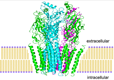
This is the nicotinic acetylcholine receptor, one of two acetylcholine receptors. It is a complex tangle of amino acids, where only molecules of a specific geometry can fit inside.

Obviously acetylcholine fits, as this is an acetylcholine receptor. This means acetylcholine is called a agonist at this receptor. Nicotine is also a agonist at the nicotinic acetylcholine receptor.

Since the structure of the receptor is so complex, other molecules can “kind of” fit, but activate the receptor only partially. Varenicline is one of these compounds, and is called a partial agonist. It simulates the pleasurable effects of nicotine, although not nearly as well, and prevents nicotine from binding to the receptors it is already attached to - which is why it is sold as an aid to quit smoking.

Other compounds can fit in the receptor, but do not activate it at all. Bupropion is one of these, and is called an antagonist. It is sold as an antidepressant and smoking cessation aid, as it binds to nicotinic acetylcholine receptors but does not activate them.

Selective Agonists

But there are two acetylcholine receptors, nicotinic and muscarinic. Acetylcholine is an agonist at both, while each compound is selective for its particular receptor.

Compound Nicotinic Acetylcholine Agonist? Muscarinic Acetylcholine Agonist?
Acetylcholine
Nicotine
Muscarine

We then call nicotine and muscarine selective agonists at the acetylcholine receptor. This concept is important in modern medicine, as a certain drug may want to target only a specific subreceptor to create a certain effect, as a full agonist may activate far too many other receptors and overwhelm the intent of the drug.

The people trying to make drugs that mimic the cancer inhibiting effects of cannabis without the pleasurable high? Yep, they’re searching for selective cannabinoid agonists, although they may be missing the point.

Neurons and Neurotransmitters

Fundamentally, the nervous system is simple. It receives sensory input, then translates this into an appropriate response. Things get a bit more complicated when we try to find out just what happens in between those two events. Sensory impulses travel through neurons, brain cells that can be thought of as switches, turning on or off depending on their input. It is important to realize that no overall response is determined solely by one structure of the brain or even one system, and it is not a simple yes-no decision but the interaction of many yes-no decisions. Like the collapse of a building or the stampede of a crowd, it is the influence of many small factors or decisions that cause a critical yet vague threshold to be reached.

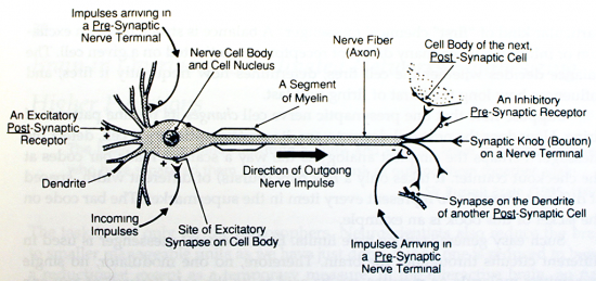
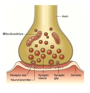
The brain can be thought of somewhat as a tangled web of neurons, with dendrites connecting to axons. At the receiving end, a single neuron thrusts out many dendrites, which bring signals toward the nerve cell. They reach out for stimuli, like branches of a tree toward the sun. Their twigs swarm with tiny receptors. Receptors are small structures designed to recognize and lock on to certain chemical compounds released by other nerve cells. The junction between these dendrites and other axons is called a synapse, and this is where the chemical release and recognition occur.

If a receptor is activated by one of these chemical signals, it adjusts the flow of ions through the membrane of the underlying nerve cell. If many receptors are activated and the flow of ions changes significantly, the nerve cell may lose its polar, charged electrical properties. Instantly the whole depolarized cell fires, sending a signal down the axon causing the release of specific chemical signals. These molecules pour out of tiny round storage packets called vesicles, and deliver this chemical message at the synapse to another dendrite. This process continually echos throughout our brain.

In general, when the first primary chemical messengers reach their receptors, they change the way the next cell acts in one of three ways.

Chemical messengers can act as transmitters. In this case the chemical messenger acts directly and is the prime mover. It quickly transfers the signal from the near side of the synapse (presynaptic side) to the far side of the synapse (postsynaptic side). To transmit its fast excitatory messages, the brain usually uses molecules of acetylcholine or glutamate. They increase the excitability of the next cell (and hence the likelyhood to fire) by depolarizing it. Other primary neurotransmitters decrease the next cells excitability. GABA, for instance, excels in this role and hyperpolarizes the next cell.

The nerve cell which released its transmitter into the synapse quickly prepares to fire again. It recaptures most of its transmitter molecules, recycles them back up into storage vesicles, and releases them again. Many nerve cells fire as fast as the wingbeat of a hummingbird, firing several hundred times a second.

We can refer to this as neurotransmission, and think of it like a private telephone line. Two people are conducting a brisk and efficient conversation, on a hardwired private link which corresponds to the synapse.

Chemical messengers can act as modulators. Instead of being the prime movers, these messengers merely modify the way other primary neurotransmitters are acting. They nudge whatever excitation or inhibition is underway in one direction or the other, but with less influence than the primary neurotransmitter. For instance, norepinephrine (NE) can act as a modulator. Some NE receptors cover the terminal endings of acetylcholine nerve cells, and can stop them from releasing their acetylcholine.

In a broad generalization, norepinephrine enhances sensate responses (including those from noxious stimuli), dopamine energizes, and serotonin enters into mood-related behaviours. Neuromodulators can be thought of as a volume knob - given an ongoing conversation, they can either amplify it or mute it.

Chemical messengers can act as neurohormones. A nerve cell may release a hormone into tissue fluids, and it may diffuse a long distance before it can find the specific receptor it binds to, and it needs no synapse to act. We can think of neurohormonal communication like a radio station broadcasting across the country. The messages travel widely, but only specific radio sets tuned into a certain station will receive any information.

The brain has trillions of these receptors, attached to dendrites, cell bodies, and axon terminals. They are precisely shaped structures that will attach to a single neurotransmitter. Each receptor can be thought of as “voting” for excitation or inhibition of the nerve cell. The result determines whether the cell fires, how frequently the cell fires, and for how long the burst of firing will last.

Each chemical messenger is used in many different circuits throughout the brain. No one modulator, no one transmitter, no one receptor can be said to be the sole cause of any behavior. A given chemical messenger does two major things:

  1. It subtly influences many behaviors by acting at many levels simultaneously in the brain.
  2. It increases or decreases the contributions made by other chemical messengers and their receptor systems.

As a result of this feedback on multiple levels, the brain is gifted with a huge array of functions and experiences, each subject to wide variation and gradation.