Posts Tagged ‘ catecholamine

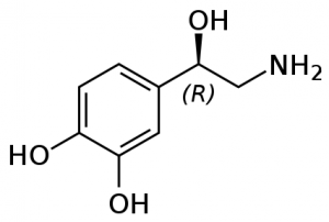
Norepinephrine

Norepinephrine (NE) is a neurotransmitter, involved in behaviours including alertness, anxiety, and attention. Most norepinephrine cells cluster in a pair of nuclei called the locus coeruleus, Latin for a “blue spot” in the brain stem. Each locus coeruleus is a long tube shaped cluster of norepinephrine neurons, both consisting of only 45,000 to 60,000 nerve cells in total. A second group of nerve cells arises deeper down in the medulla, and delivers norepinephrine mostly to the hypothalamus.

Primate brains have a dense network supplying norepinephrine to the posterior parietal lobe (which integrates sensory information), to part of the thalamus, and to the outer layers of the optic lobes in the midbrain. Relatively little norepinephrine is supplied to the temporal cortex, involved in speech, high-level visual processing (ie facial recognition), and memory. This outlines a distinctive feature of norepinephrine, that it is generally involved in direct sensory input rather than sensory processing. But the hypothalamus (which regulates hunger, thirst, fatigue, and sleep cycles) has the highest concentration of norepinephrine at 1150 nanograms per gram, indicating the vital role this neurotransmitter plays in deep primal instincts.

NE cells in the locus coeruleus fire especially in response to stressful, painful, noxious stimuli. In rats, they fire faster for up to thirty seconds if the toe is compressed in a painful manner. A strong but generic stimuli for cats (say a loud noise) will prompt a brief discharge of norepinephrine, but this response dwindles as the stimuli is repeated and the cat becomes conditioned.

In higher primates like monkeys, pain and stress also cause norepinephrine cells to fire faster. But what really gets monkey norepinephrine production going is fruit juice, their favorite treat. Norepinephrine cells fire at their highest rates when drinking fruit juice, seven to fifteen times per second. The norepinephrine cells of primate brains appear to be slightly different than that of lower animals, as they are especially activated by noxious stimuli and also certain specific attractive stimuli. Norepinephrine cells fire much slower if primates are given opiate based painkillers however, and during dream states (REM) of sleep.

So what exactly happens when norepinephrine cells fire more frequently? They appear to have rather few direct effects, but act primarily as a modulator. For instance, norepinephrine alone does little to change the slow background firing of cells in the hippocampus proper (involved in memory and spatial navigation) - but if these cells have been excited by addition of glutamate, norepinephrine makes them fire even faster. Norepinephrine also makes hippocampal cells fire faster if they have been excited by natural outside visual and auditory stimuli. It also reduces spontaneous background activity, increasing the signal to noise ratio of sensation from outside. This increased signal to noise ratio has become very valuable in treating symptoms of conditions such as ADHD. Norepinephrine and dopamine both play a large role in attention and focus. Various amphetamines and other compounds like methylphenidate (Ritalin) prescribed for ADHD cause higher levels of norepinephrine and dopamine.

Other compounds can act as agonists at norepinephrine receptors. Clonidine is used to relieve the suffering of addicts withdrawing from opiates. It activates certain autoreceptors which slow the norepinephrine cells’ firing rate, reducing symptoms such as anxiety, restlessness, and aching in muscles and joints. This clinical success emphasizes the theories linking excessive norepinephrine activity within the locus coeruleus with states of anxiety and tension. For instance, investigation into patients suffering panic attacks suggests that they may lack the normal mechanisms that would hold their norepinephrine systems in check.
Dopamine

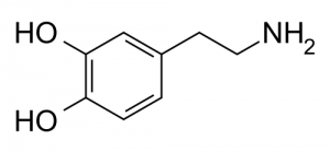
Dopamine (DA) is a neurotransmitter present in many animals, from the honeybee to the dolphin. It is involved in many behaviors associated with motivation and reward. The majority of dopamine in humans is released from two areas.

First is the the substantia nigra, whose darkly pigmented cell bodies supply dopamine to the deep motoric nuclei of the basal ganglia. Dopamine concentrations in one nuclei, the putamen, exceed anywhere else in the brain reaching a level of 5740 nanograms per gram. Dopamine released in this nigrostriatal pathway helps us quickly execute our motor patterns and associated movements. For instance, if dopamine drops to very low levels on one side of this system Parkinsonian symptoms will arise, with sluggish arm and leg movements on one side of the body.

Second is the ventral tegmental system. Its cells send much of their dopamine up to the nucleus accumbens in the ventral striatum, and a separate pathway supplies the limbic system. Two other branches supply dopamine to the cortex in the prefrontal and cingulate regions. An especially dense network of dopamine fibers covers the inner part of the prefrontal lobe, the same region where the thalamus sends its major input. The intersection of these two pathways in the prefrontal cortex remains one of the brain’s most constant features, dating back to the tree shrew and reminding us of our origins in the forest.

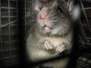
Suppose you take a rat, normally a social animal, and reduce its social stimuli over a prolonged period making it a hermit in a cage. The different dopamine systems react in different ways - the metabolic activity of the dopamine cells supplying the frontal lobes slows, whereas metabolism increases in dopamine cells projecting up to the dorsal and ventral striatum. Even though the isolated rats have now quieted down and show few spontaneous behaviors (frontal lobes), they jump more when an electrical shock is delivered to the foot (dorsal and ventral striatum). Increased social exposure causes a reversal of these effects - more active and less nervous rats.

Each person’s brain is unique, as the number and distribution of dopamine receptors varies widely. Early in life, brain development responds to male or female hormones present. Male rats already show more evidence of dopamine receptors in their cortex and amygdala only a few days after being born.

There are five known dopamine subreceptors in humans. The most abundant dopamine subreceptor in the nervous system is the D1 receptor, which regulate neuronal growth and development and mediates some behavioral responses. It also stimulates adenylyl cyclase and activates cyclic AMP-dependent protein kinases, involved in the regulation of glycogen, sugar, and lipid metabolism. D2 receptors are gaining prominence as potential keys to certain diseases, as increased D2 receptor levels have been linked to schizophrenia, certain Parkinson’s symptoms, and narcolepsy.

The major functional role of dopamine systems in the human brain appears twofold. First, there are prominent motoric effects. Decades of research on rats and mice have shown that any cause of indirect release of dopamine into the nucleus accumbens causes rats to engage in specific motor sequences: they move around more, they sniff more, and engage in repetitive grooming behaviors. This is not unlike a human who has experienced dopamine increase caused by cocaine or other drug consumption, as they fidget, sniff and lick their lips, run their fingers through their hair, or pick at the skin.

But there is more than just a general mobilizing or energizing effect. This became clear when experiments were undertaken as animals pressed on a lever, working to receive food as a reward. Drugs which increased dopamine levels caused the animals to become more selective and efficient, suggesting that dopamine helps to sustain goal directed behavior. While dopamine was initially dubbed the “pleasure” neurotransmitter, recent research suggests that pleasure is a less appropriate term than “motivation”. As any speed freak will tell you, after a certain point self-administration of the drug becomes distinctly dysphoric, but the desire to continue dosing remains strong and is not correlated with the level of
“pleasure” (if any) that the next dose will produce.

There are also interesting correlations to personality. “Extroverted” personalities tend to show higher spinal fluid levels of dopamine breakdown products, and a set of aggressive patients also showed the same evidence of higher dopamine turnover. After practicing yoga meditation for six months however, the levels of dopamine breakdown products of the aggressive patients fell from an average of 51 nanograms per liter to 41 nanograms per liter.