Archive for the ‘ Physiology ’ Category

Form Constants and the Visual Cortex

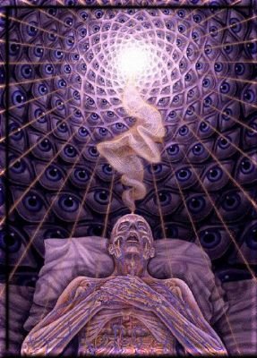
There are common visual concepts which cut across boundaries of culture and time and reflect what it truly means to be human. Near death experiences are often associated with seeing a “light at the end of a tunnel”. In the Bible, God appeared to Ezekiel as a “wheel within a wheel”. Spirals and concentric circles are commonly found in petrogylphs carved by cultures long dead. Similar visual effects are reported during extreme psychological stress, fever delirium, psychotic episodes, sensory deprivation, and are reliably induced by psychedelic drugs.

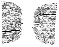
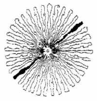
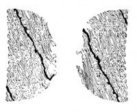
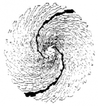
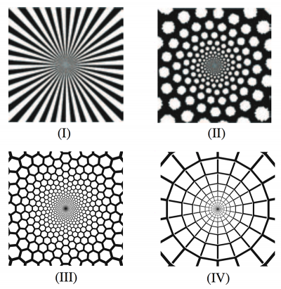
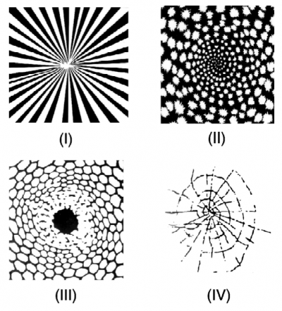
In 1926, Heinrich Klüver undertook a groundbreaking series of experiments where he categorized the visual effects produced by mescaline. Various volunteers were recruited, peyote administered, reports taken, and results classified into categories. There were general perceptual effects, variations in color and distortions of shape. But the most interesting reports were consistent visual concepts he dubbed “form constants”. Across many volunteers and many sessions, all reported seeing visual patterns with similar structure.

They were classified into four main types: I) tunnels, II) spirals, III) lattices, and IV) cobwebs. For almost fifty years, these form constants were regarded as a strange mystery of visual perception, a seemingly unexplainable common human experience.

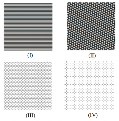
In 1979 Jack Cowan and G. Bard Ermentrout put forward a very interesting explanation, supported by a rigorous mathematical treatment. These visual effects are the result of specific noise patterns in the visual cortex, which are then transformed by the wiring between the brain and the eye to produce these unique shapes. They generated simple biologically allowable noise patterns, transformed them, and produced graphs of these form constants.

Let’s dive a bit deeper into how this was done, by first having a look at the structure of the visual cortex. We’ll look specifically at V1, the first layer of visual processing where information from the retina is fed to. We can think of it as a sheet of hypercolumns, cells sensitive to lines oriented in any direction. This surface is crinkled up like a ball of paper in your brain, but we can unfold it in a theoretical sense. These hypercolumns are linked together in a specific manner, which allows noise patterns of only certain types to form. Just like a tarp in the wind will only flap in certain predictable ways assuming it does not tear, so too will noise only travel across the visual cortex in specific ways. Four types of noise were found.

These stable planforms can be thought of as excited noisy states in contrast to the normal low-activity state of the visual cortex. We can refer to them as I) the non-contoured roll, II) the non-contoured hexagon, III) the even contoured hexagon, and IV) the even contoured square.

So now that we have our noise patterns, how are they mapped from the visual cortex to what we actually see? Biology provides a clue here. Experiments have been done which allow mapping of how the visual cortex represents input from the retina. By stimulating a certain point or region of the retina, the corresponding cells which light up in V1 can be measured. The easiest mapping you might think of would be for the input of the retina to be represented as a flat sheet, which is then passed to the visual cortex like a photocopy. Instead, it turns out that the circular retina’s image is twisted and mapped in a slightly more complex manner to the flat surface of V1.

Visual Cortex (V1) Retina

We can see that straight lines in our visual cortex are mapped to curved lines in the retina, and vice versa. We can represent this relationship mathematically using the complex logarithm, so let’s apply this complex log transform to our four types of visual cortex noise.

And beautifully, Klüver’s four form constants are produced, visual cortex noise twisted by the wiring between mind and eye. This hypothesis fits the fact that these high energy states may be caused by a variety of stimuli affecting excitability of the brain but most reliably by psychedelic drugs which bind to serotonin receptors richly expressed in the visual cortex.

It is compelling to think that these powerful symbols rely on no religion, no culture, and no time. They are a product of the fact that we are all human and share the same biology. A true tragedy that these visions have been used as an excuse to kill others when we all see the same wheels within wheels.

Ermentrout, G.B. and Cowan, J.D., “A mathematical theory of visual hallucination patterns.” Biol. Cybernet. 34 (1979), no. 3, 137-150.

Bressloff, Paul C.; Cowan, Jack D.; Golubitsky, Martin; Thomas, Peter J.; Weiner, Matthew C. (March 2002). “What Geometric Visual Hallucinations Tell Us About the Visual Cortex“. Neural Computation (The MIT Press) 14 (3): 473–491.
DMT and the Pineal Gland

One of the most popular “drug geek” myths is that the powerful psychedelic compound DMT is produced naturally within your body, specifically the pineal gland. Not only this, but this natural DMT is apparently involved in a wide variety of previously unexplained processes - it is the mechanism of dreaming, it causes religious feelings, and DMT production spikes near death to “carry away the soul”. This appears to stem primarily from Dr. Richard Strassman’s book The Spirit Molecule, which advanced many of these hypotheses which were then passed on and extrapolated telephone-game style to the point where fluoridated water is apparently an Illuminati plot to suppress natural DMT production.

There’s just one problem - there doesn’t appear to be any concrete evidence whatsoever for this. Dr. Strassman himself explains:

I did my best in the DMT book to differentiate between what is known, and what I was conjecturing about (based upon what is known), regarding certain aspects of DMT dynamics. However, it’s amazing how ineffective my efforts seem to have been. So many people write me, or write elsewhere, about DMT, and the pineal, assuming that the things I conjecture about are true. When I was writing the book, I thought I was clear enough, and repeating myself would have gotten tedious.

We don’t know whether DMT is made in the pineal. I muster a lot of circumstantial evidence supporting a reason to look long and hard at the pineal, but we do not yet know. There are data suggesting urinary DMT rises in psychotic patients when their psychosis is worse. However, we don’t know whether DMT rises during dreams, meditation, near-death, death, birth or any other endogenous altered state. To the extent those states resemble those brought on by giving DMT, it certainly makes one wonder if endogenous DMT might be involved, and if it were, it would explain a lot. But we don’t know yet. Even if the pineal weren’t involved, that would have little overall effect on my theories regarding a role for DMT in endogenous altered states, because we do know that the gene involved in DMT synthesis is present in many organs, particularly lung. If the pineal made DMT, it would tie up a lot of loose ends regarding this enigmatic little organ. But people seem to live pretty normals lives without a pineal gland; for example, when it has had to be removed because of a tumor.

In both these regards-the pineal-DMT connection, and endogenous DMT dynamics-we ought to know a lot more within the next several years due to the efforts of a research group being led by Steven Barker at Louisiana State University. He, with his grad student Ethan McIlhenny, are developing a new super-assay for DMT, 5-MeO-DMT, bufotenine, and metabolites. This assay will be capable of detecting those compounds much more sensitively than previous generations of assays. They’re looking at endogenous levels in awake sober normals, to assess baseline values of these compounds. We should have some data from those samples within a year. They also will be looking at pineal tissue. Once we have some baseline data in normal humans in normal waking consciousness, comparisons can be made between those levels and levels in endogenous altered states, like dreams, near-death, and so on.

It appears that myths about drugs can cut both ways, and this is an important illustration of the requirement for critical thinking, no matter how appealing the initial conjecture. Steven Barker and Ethan McIlhenny’s work to determine baseline DMT levels continues, with their latest paper involving detection of metabolites produced by ayahuasca consumption.

Hanna J. “DMT and the Pineal: Fact or Fiction?” www.erowid.org/chemicals/dmt/dmt_article2.shtml. Jun 3 2010.

Receptors and Agonism

We know that receptors in a synapse bind to neurotransmitters, and this binding can change the likelyhood of a nerve cell to fire. But do these receptors only bind to one specific neurotransmitter, or is there more to it? We can think of a receptor as a lock that only a specific key can open, a particular geometric and chemical structure that only certain molecules can interact with.

Agonists, Partial Agonists, and Antagonists

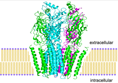
This is the nicotinic acetylcholine receptor, one of two acetylcholine receptors. It is a complex tangle of amino acids, where only molecules of a specific geometry can fit inside.

Obviously acetylcholine fits, as this is an acetylcholine receptor. This means acetylcholine is called a agonist at this receptor. Nicotine is also a agonist at the nicotinic acetylcholine receptor.

Since the structure of the receptor is so complex, other molecules can “kind of” fit, but activate the receptor only partially. Varenicline is one of these compounds, and is called a partial agonist. It simulates the pleasurable effects of nicotine, although not nearly as well, and prevents nicotine from binding to the receptors it is already attached to - which is why it is sold as an aid to quit smoking.

Other compounds can fit in the receptor, but do not activate it at all. Bupropion is one of these, and is called an antagonist. It is sold as an antidepressant and smoking cessation aid, as it binds to nicotinic acetylcholine receptors but does not activate them.

Selective Agonists

But there are two acetylcholine receptors, nicotinic and muscarinic. Acetylcholine is an agonist at both, while each compound is selective for its particular receptor.

Compound Nicotinic Acetylcholine Agonist? Muscarinic Acetylcholine Agonist?
Acetylcholine
Nicotine
Muscarine

We then call nicotine and muscarine selective agonists at the acetylcholine receptor. This concept is important in modern medicine, as a certain drug may want to target only a specific subreceptor to create a certain effect, as a full agonist may activate far too many other receptors and overwhelm the intent of the drug.

The people trying to make drugs that mimic the cancer inhibiting effects of cannabis without the pleasurable high? Yep, they’re searching for selective cannabinoid agonists, although they may be missing the point.

Neurons and Neurotransmitters

Fundamentally, the nervous system is simple. It receives sensory input, then translates this into an appropriate response. Things get a bit more complicated when we try to find out just what happens in between those two events. Sensory impulses travel through neurons, brain cells that can be thought of as switches, turning on or off depending on their input. It is important to realize that no overall response is determined solely by one structure of the brain or even one system, and it is not a simple yes-no decision but the interaction of many yes-no decisions. Like the collapse of a building or the stampede of a crowd, it is the influence of many small factors or decisions that cause a critical yet vague threshold to be reached.

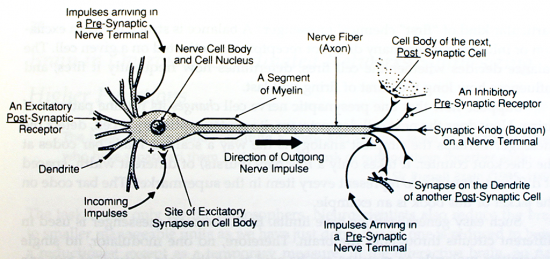
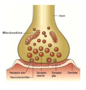
The brain can be thought of somewhat as a tangled web of neurons, with dendrites connecting to axons. At the receiving end, a single neuron thrusts out many dendrites, which bring signals toward the nerve cell. They reach out for stimuli, like branches of a tree toward the sun. Their twigs swarm with tiny receptors. Receptors are small structures designed to recognize and lock on to certain chemical compounds released by other nerve cells. The junction between these dendrites and other axons is called a synapse, and this is where the chemical release and recognition occur.

If a receptor is activated by one of these chemical signals, it adjusts the flow of ions through the membrane of the underlying nerve cell. If many receptors are activated and the flow of ions changes significantly, the nerve cell may lose its polar, charged electrical properties. Instantly the whole depolarized cell fires, sending a signal down the axon causing the release of specific chemical signals. These molecules pour out of tiny round storage packets called vesicles, and deliver this chemical message at the synapse to another dendrite. This process continually echos throughout our brain.

In general, when the first primary chemical messengers reach their receptors, they change the way the next cell acts in one of three ways.

Chemical messengers can act as transmitters. In this case the chemical messenger acts directly and is the prime mover. It quickly transfers the signal from the near side of the synapse (presynaptic side) to the far side of the synapse (postsynaptic side). To transmit its fast excitatory messages, the brain usually uses molecules of acetylcholine or glutamate. They increase the excitability of the next cell (and hence the likelyhood to fire) by depolarizing it. Other primary neurotransmitters decrease the next cells excitability. GABA, for instance, excels in this role and hyperpolarizes the next cell.

The nerve cell which released its transmitter into the synapse quickly prepares to fire again. It recaptures most of its transmitter molecules, recycles them back up into storage vesicles, and releases them again. Many nerve cells fire as fast as the wingbeat of a hummingbird, firing several hundred times a second.

We can refer to this as neurotransmission, and think of it like a private telephone line. Two people are conducting a brisk and efficient conversation, on a hardwired private link which corresponds to the synapse.

Chemical messengers can act as modulators. Instead of being the prime movers, these messengers merely modify the way other primary neurotransmitters are acting. They nudge whatever excitation or inhibition is underway in one direction or the other, but with less influence than the primary neurotransmitter. For instance, norepinephrine (NE) can act as a modulator. Some NE receptors cover the terminal endings of acetylcholine nerve cells, and can stop them from releasing their acetylcholine.

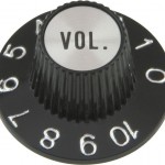
In a broad generalization, norepinephrine enhances sensate responses (including those from noxious stimuli), dopamine energizes, and serotonin enters into mood-related behaviours. Neuromodulators can be thought of as a volume knob - given an ongoing conversation, they can either amplify it or mute it.

Chemical messengers can act as neurohormones. A nerve cell may release a hormone into tissue fluids, and it may diffuse a long distance before it can find the specific receptor it binds to, and it needs no synapse to act. We can think of neurohormonal communication like a radio station broadcasting across the country. The messages travel widely, but only specific radio sets tuned into a certain station will receive any information.

The brain has trillions of these receptors, attached to dendrites, cell bodies, and axon terminals. They are precisely shaped structures that will attach to a single neurotransmitter. Each receptor can be thought of as “voting” for excitation or inhibition of the nerve cell. The result determines whether the cell fires, how frequently the cell fires, and for how long the burst of firing will last.

Each chemical messenger is used in many different circuits throughout the brain. No one modulator, no one transmitter, no one receptor can be said to be the sole cause of any behavior. A given chemical messenger does two major things:

  1. It subtly influences many behaviors by acting at many levels simultaneously in the brain.
  2. It increases or decreases the contributions made by other chemical messengers and their receptor systems.

As a result of this feedback on multiple levels, the brain is gifted with a huge array of functions and experiences, each subject to wide variation and gradation.

Videos of the Brain

Some great videos illustrating brain anatomy. First, anatomy and function using color coded 3D animation.


Next, a more in depth dissection with Ami Cohen, a graduate student at USCB.


The Inside of the Brain

Now let’s work our way inside the brain.

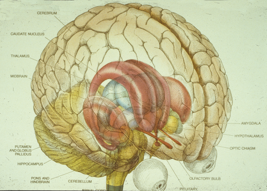
At the base of the brain the spinal cord becomes enlarged, and is known as the brain stem. The lowest level of the brain stem is the medulla, rising up from the spinal cord. The medulla controls the most basic of operations including cardiac, respiratory, vomiting and vasomotor centers, as well as regulating autonomic functions, such as breathing, heart rate and blood pressure.

As we move further up the brain stem we find the pons, involved in general arousal of the nervous system, assisting in controlling autonomic functions, sleep, and relaying sensory information between the cerebrum and cerebellum. This relaying is necessary as the cerebellum is seemingly detached from the rest of the brain, lying off to the back of the brain stem. It is involved in other basic functions, most notably motor control. It does not initiate movement, but acts as a grand director - receiving input from sensory systems and from other parts of the brain and spinal cord, and integrating these inputs to fine tune gross motor activity. The cerebellum is also involved in motor learning, such as a baby deer tottering around and correlating sensory input to motor output in order to use its legs.

The midbrain controls the visual and auditory systems as well as eye movement. Portions of the midbrain called the red nucleus and the substantia nigra are involved in the control of body movement. The darkly pigmented substantia nigra contains a large number of dopamine-producing neurons, degeneration of which is associated with Parkinson’s disease. Barry Kidston, a 23-year-old chemistry graduate student in Maryland, synthesized MPPP (a synthetic opioid painkiller) in 1976 after learning about it from an obscure 1947 paper, but screwed up a few tiny technical details leaving traces of MPTP behind. It took only three days of self-injection of the tainted MPPP for the trace MPTP to irreversibly damage the substantia nigra and for Barry to start exhibiting symptoms of Parkinson’s disease.

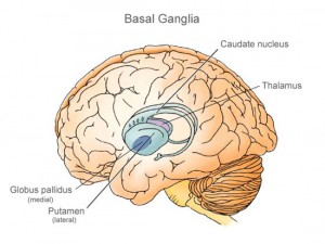
The bulbous thalmus extends out of the top of the brain stem, and it situated between the cerebral cortex and the midbrain, both in terms of location and function. Its functions include relaying sensory input and motor signals to the cerebral cortex, along with the regulation of consciousness, sleep and alertness. It is surrounded by basal ganglia which are primarily involved in action selection, or the decision of which of several possible behaviors to execute at a given time. Experimental studies show that the basal ganglia exert an inhibitory influence on a number of motor systems, and that a release of this inhibition permits a motor system to become active. The basal ganglia can therefore be thought of as a “behaviour switch” influenced by signals from many parts of the brain, including the “executive” prefrontal cortex.

Centered deep below the thalamus lies the hypothalamus, the connection between the nervous system and the endocrine (hormonal) system. The hypothalamus controls body temperature, hunger, thirst, fatigue, and circadian cycles. Sweeping up from the hypothalamus on both sides are several other structures collectively known as the limbic system, or “paleomammalian brain”. These ancient structures infuse emotional overtones in our experience and into our affective responses, affect long term memory, behaviour, and the sense of smell. One limbic component, the hippocampus, helps us process data and relay them to be stored in various memory circuits.

The limbic system was originally proposed as the emotional center of the brain, with the neocortex taking the role of “hard computation”. This has been cast into controversy with the discovery that the hippocampus is involved in memory retention, and the boundaries of the limbic system have been redrawn again and again since the concept was originally proposed. Some scientists propose scrapping the concept of a seperate limbic system entirely.

The Outside of the Brain

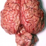
The brain is divided into two hemispheres, one on the left and one on the right, connected by a thick band of tissue called the corpus callosum. Oddly enough, the right hemisphere controls the left side of the body, and the left hemisphere controls the right side of the body. Activity in the left hemisphere is correlated with speech, writing, language, and calculation. Activity in the right hemisphere is correlated with spatial abilities, face recognition, and some musical abilities.

The outside of the brain is a grey layer, folded into strange ridges, and called the cerebral cortex, or the “gray matter”. The gray color comes from billions of nerve cells, and connects to bundles of of white, insulated fibers - “white matter”. You can think of it like an outer “rind” - in humans, it makes up 80% of the brain. In monkeys, only about 66%. The amount of “gray matter” appears to contribute significantly to how advanced a species is, and humans have the most of it.

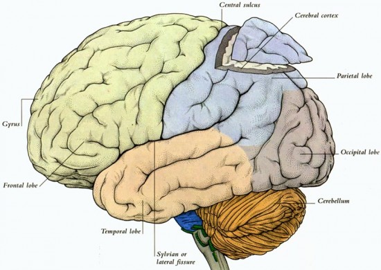
The brain itself can be divided roughly into four lobes.

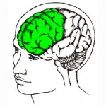
The frontal lobes help make us human. The association cortex in this region helps us generate goals that are personally desirable, to determine how socially appropriate they are, and then to decide which behaviors will result in the best future outcome. Many higher order executive functions arise in the frontal lobes, including those that direct speech and other precisely managed movements. The frontal lobe contains most of the dopamine-sensitive neurons in the cerebral cortex.
Behind lie the parietal lobes. These lobes not only receive sensation but allow fine discrimination between them. When we fumble in our pocket, which coin is a nickel and which coin is a quarter? Our fingers tells us “this one”, as the parietal lobes make abstract representations of the sensations from our fingers, relating these representations to the type of coin.
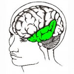
Below each parietal lobe, deep to the temple on either side, are the temporal lobes. They decode and interpret what we hear and see, and process other more elaborate, patterned sensory messages. For instance, the left temporal lobe is important in understanding language-related concepts.
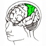
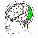
The occipital lobes at the back of the brain register impulses concerned with vision. They then pattern them into streams of visual messages that are relayed forward to both the temporal and parietal lobes. Note that they do not relay patterns of photons hitting your eye, but more abstract and compressed representations. These mental images are generated and regenerated, and could be thought of as our “mind’s eye”, the mental templates that allow us to recognize and insert meaning into what we see. Strong increases in occipital activity is noted under the influence of certain psychedelic drugs.