Archive for the ‘ Neurotransmitters ’ Category

Endocannabinoids

The precise mode of action of cannabis was unclear until very recently. In 1988 cannabinoid receptors were shown to exist in a rat brain, and in 1990 the locations of the cannabinoid receptor system were mapped in several mammalian species, including man. This led to the search for endogenous cannabinoids (endocannabinoids), natural compounds synthesized within the brain that would activate these same receptors. Several were discovered, with anandamide and 2-AG receiving the most attention. It was found that endocannabinoids operate via retrograde signalling, where a signal travels in reverse from a postsynaptic neuron to a presynaptic one. Unlike other neurotransmitters such as the monoamines which are stored in vesicles for eventual release after synthesis, endocannabinoids are created “on demand” by enzymes as required.
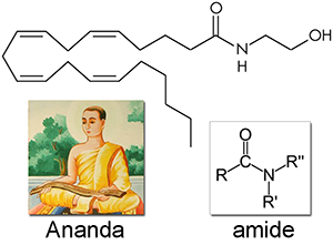
Anandamide (AEA) was first isolated in 1992 and named after ananda, the Sanskrit word for bliss, and the amide chemical backbone. Like all of the endocannabinoids, it is made from arachidonic acid in our bodies, an essential polyunsaturated omega-6 fatty acid that must be consumed as part of a complete diet. Anandamide is a full agonist at CB1 receptors with a potency comparable to THC, and a partial agonist at CB2 receptors. It is very common, found in nearly all tissues and in a wide range of animals.
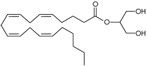
2-arachidonoyl glycerol (2-AG). No one really wants to admit it since anandamide was discovered first and has such a great name, but 2-arachidonoyl glycerol is probably chiefly responsible for endocannabinoid signaling. A full agonist at both CB1 and CB2 receptors, it is capable of stimulating higher G-protein activation and is present at significantly higher concentrations in the brain than anandamide.
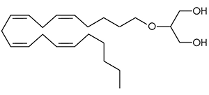
2-arachidonyl glyceryl ether (noladin ether) was first discovered in the brain tissue of a pig in 2001, and prior to this had been synthesized in a lab as a closely related analog of 2-AG. It binds primarily to the CB1 receptor with weak agonism at the CB2 receptor, and is more metabolically stable than 2-AG resulting in a longer half life.
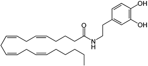
N-arachidonoyl-dopamine (NADA) Discovered in 2000 in the brain of rats, NADA preferentially binds to the CB1 receptor with no action at dopamine receptors. It the amide of the neurotransmitter dopamine and arachidonic acid, as well as a potent inhibitor of the growth of breast cancer cells.
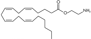
Virodhamine (OAE) can be thought of as similar in structure to anandamide where the nitrogen and oxygen atoms have switched places. The molecule was therefore named virodhamine after the Sanskrit word virodha meaning opposite. It is a partial agonist at the CB1 receptor and a full agonist at the CB2 receptor. It is present in concentrations comparable to anandamide in the hippocampus, but is present much higher concentrations in peripheral tissues with CB2 receptors.
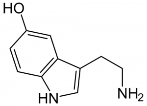

Serotonin

Serotonin (5-hydroxytryptamine, ST, 5HT) is a hydroxylated tryptamine, a class of compounds which rose to widespread fame in the psychedelic drug community with the publication of Alexander Shulgin’s work “Tryptamines I Have Known and Loved“. Most of the body’s serotonin (80%+) is produced and contained in the digestive tract, where it regulates intestinal movements. The remainder is synthesized in Raphe cells deep along the midline core of the brainstem, where it is involved in activities such as mood, appetite, sleep, muscle contraction, memory, and learning. Raphe cells start to fire slowly, and their firing also tails off slowly. This reflects a system designed to modulate slower state-dependent activities, rather than a quick response to external stimuli.

Serotonin is present in all bilaterally symmetric animals, and acts to modulate digestive movements and is involved in the perception of resource availability. For very simple animals resources could only mean food, but in more advanced animals such as primates this also includes social status (or lack of it). Serotonin can modulate an animal’s rate of growth, desire to reproduce, and overall mood in relation to the perceived abundance or scarcity of these resources. In roundworms serotonin is released as a signal in response to positive events such as finding a new grazing ground or meeting a suitable mate. Lobsters injected with serotonin begin to display dominant social behavior, regardless of previous social status.

There are at least three major families of serotonin receptors. Many psychedelic drugs, such as psilocybin, act as partial serotonin agonists primarily active at the 5HT2 receptor. But there is some action at additional receptors, including not surprisingly the 5HT3 receptor involved in feelings of nausea. 5HT3 antagonists such as ondansetron are used during chemotherapy and are considered the gold standard in anti-nausea treatment.

In primates, many serotonin terminals end in the fourth layer of the visual cortex, where they can influence visual perception. Additionally, the temporal lobe is rich in serotonin nerve cell endings, meaning serotonin action has a significant influence on the stream of visual interpretations and association arising from external stimuli. Serotonin tends to act generally as an inhibitor in the cortex, where it reduces the excitatory responses evoked by other sensory stimuli. It also tends to preserve the general noise level of firing in the background, reducing the signal to noise ratio in contrast to the action of norepinephrine.

In humans, low serotonin levels are correlated with certain states of depression. This is reinforced by the fact that consumption of the serotonin precursor 5-hydroxytryptophan (5HTP) causes alleviation of depressive symptoms in some patients. The brain normally has a dense concentration of postsynaptic 5HT2 receptors in the prefrontal cortex, the location in the brain thought to play an “executive” role in coordinating sensory response. Patients who have had a major depressive incident tend to have even higher numbers of these receptors, which is thought to be a result of the brain compensating for lower serotonin levels. In the brain stems of depressed subjects, autopsies have also shown reduced serotonin levels.

Increases in serotonin levels tend to have the opposite effect, increasing outward social behaviours. In monkeys, serotonin agonist drugs increase the approaching, grooming, resting, and eating activities typical of the dominant males. They also reduce inward social solitary, vigilant, and avoidance behaviors. This is reflected in humans under the influence of MDMA, an amphetamine derivative which simultaneously releases large amounts of serotonin while blocking its reuptake. Subjects become much more social and talkative, empathy towards others is increased, and anxiety and paranoia disappear. This has made MDMA and related compounds ideal for psychiatric therapy, particularly for couples and those who have experienced intensely traumatic events.

But the “serotonin as happiness” message sold by drug companies pushing antidepressants is not a complete story. A depressed person may have decreased serotonin levels in response to environmental or other factors, and an antidepressant will certainly raise them. But if these stress factors do not change along with the serotonin levels (through changes in behavior or the environment itself), it is unlikely to create lasting positive change. Serotonin appears to act to regulate the extent or intensity of moods, with less influence on the direct attitude of the mood itself. Similar to the use of MDMA in therapy, these drugs should be regarded as a catalyst to move in the correct direction, and not a magic bullet.

Norepinephrine

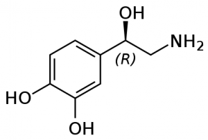
Norepinephrine (NE) is a neurotransmitter, involved in behaviours including alertness, anxiety, and attention. Most norepinephrine cells cluster in a pair of nuclei called the locus coeruleus, Latin for a “blue spot” in the brain stem. Each locus coeruleus is a long tube shaped cluster of norepinephrine neurons, both consisting of only 45,000 to 60,000 nerve cells in total. A second group of nerve cells arises deeper down in the medulla, and delivers norepinephrine mostly to the hypothalamus.

Primate brains have a dense network supplying norepinephrine to the posterior parietal lobe (which integrates sensory information), to part of the thalamus, and to the outer layers of the optic lobes in the midbrain. Relatively little norepinephrine is supplied to the temporal cortex, involved in speech, high-level visual processing (ie facial recognition), and memory. This outlines a distinctive feature of norepinephrine, that it is generally involved in direct sensory input rather than sensory processing. But the hypothalamus (which regulates hunger, thirst, fatigue, and sleep cycles) has the highest concentration of norepinephrine at 1150 nanograms per gram, indicating the vital role this neurotransmitter plays in deep primal instincts.

NE cells in the locus coeruleus fire especially in response to stressful, painful, noxious stimuli. In rats, they fire faster for up to thirty seconds if the toe is compressed in a painful manner. A strong but generic stimuli for cats (say a loud noise) will prompt a brief discharge of norepinephrine, but this response dwindles as the stimuli is repeated and the cat becomes conditioned.

In higher primates like monkeys, pain and stress also cause norepinephrine cells to fire faster. But what really gets monkey norepinephrine production going is fruit juice, their favorite treat. Norepinephrine cells fire at their highest rates when drinking fruit juice, seven to fifteen times per second. The norepinephrine cells of primate brains appear to be slightly different than that of lower animals, as they are especially activated by noxious stimuli and also certain specific attractive stimuli. Norepinephrine cells fire much slower if primates are given opiate based painkillers however, and during dream states (REM) of sleep.

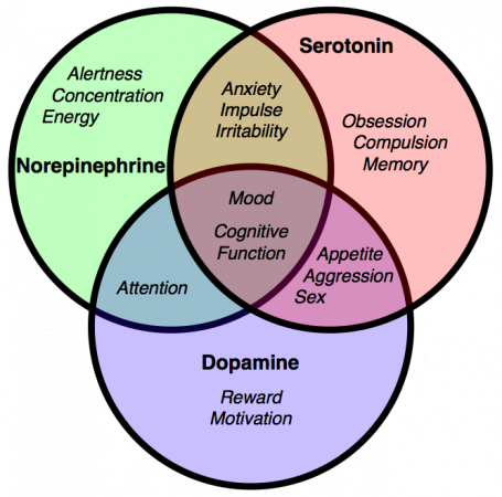
So what exactly happens when norepinephrine cells fire more frequently? They appear to have rather few direct effects, but act primarily as a modulator. For instance, norepinephrine alone does little to change the slow background firing of cells in the hippocampus proper (involved in memory and spatial navigation) - but if these cells have been excited by addition of glutamate, norepinephrine makes them fire even faster. Norepinephrine also makes hippocampal cells fire faster if they have been excited by natural outside visual and auditory stimuli. It also reduces spontaneous background activity, increasing the signal to noise ratio of sensation from outside. This increased signal to noise ratio has become very valuable in treating symptoms of conditions such as ADHD. Norepinephrine and dopamine both play a large role in attention and focus. Various amphetamines and other compounds like methylphenidate (Ritalin) prescribed for ADHD cause higher levels of norepinephrine and dopamine.

Other compounds can act as agonists at norepinephrine receptors. Clonidine is used to relieve the suffering of addicts withdrawing from opiates. It activates certain autoreceptors which slow the norepinephrine cells’ firing rate, reducing symptoms such as anxiety, restlessness, and aching in muscles and joints. This clinical success emphasizes the theories linking excessive norepinephrine activity within the locus coeruleus with states of anxiety and tension. For instance, investigation into patients suffering panic attacks suggests that they may lack the normal mechanisms that would hold their norepinephrine systems in check.

Dopamine

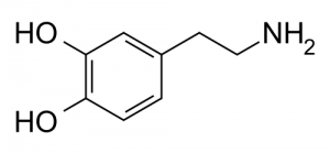
Dopamine (DA) is a neurotransmitter present in many animals, from the honeybee to the dolphin. It is involved in many behaviors associated with motivation and reward. The majority of dopamine in humans is released from two areas.

First is the the substantia nigra, whose darkly pigmented cell bodies supply dopamine to the deep motoric nuclei of the basal ganglia. Dopamine concentrations in one nuclei, the putamen, exceed anywhere else in the brain reaching a level of 5740 nanograms per gram. Dopamine released in this nigrostriatal pathway helps us quickly execute our motor patterns and associated movements. For instance, if dopamine drops to very low levels on one side of this system Parkinsonian symptoms will arise, with sluggish arm and leg movements on one side of the body.

Second is the ventral tegmental system. Its cells send much of their dopamine up to the nucleus accumbens in the ventral striatum, and a separate pathway supplies the limbic system. Two other branches supply dopamine to the cortex in the prefrontal and cingulate regions. An especially dense network of dopamine fibers covers the inner part of the prefrontal lobe, the same region where the thalamus sends its major input. The intersection of these two pathways in the prefrontal cortex remains one of the brain’s most constant features, dating back to the tree shrew and reminding us of our origins in the forest.

Suppose you take a rat, normally a social animal, and reduce its social stimuli over a prolonged period making it a hermit in a cage. The different dopamine systems react in different ways - the metabolic activity of the dopamine cells supplying the frontal lobes slows, whereas metabolism increases in dopamine cells projecting up to the dorsal and ventral striatum. Even though the isolated rats have now quieted down and show few spontaneous behaviors (frontal lobes), they jump more when an electrical shock is delivered to the foot (dorsal and ventral striatum). Increased social exposure causes a reversal of these effects - more active and less nervous rats.

Each person’s brain is unique, as the number and distribution of dopamine receptors varies widely. Early in life, brain development responds to male or female hormones present. Male rats already show more evidence of dopamine receptors in their cortex and amygdala only a few days after being born.

There are five known dopamine subreceptors in humans. The most abundant dopamine subreceptor in the nervous system is the D1 receptor, which regulate neuronal growth and development and mediates some behavioral responses. It also stimulates adenylyl cyclase and activates cyclic AMP-dependent protein kinases, involved in the regulation of glycogen, sugar, and lipid metabolism. D2 receptors are gaining prominence as potential keys to certain diseases, as increased D2 receptor levels have been linked to schizophrenia, certain Parkinson’s symptoms, and narcolepsy.

The major functional role of dopamine systems in the human brain appears twofold. First, there are prominent motoric effects. Decades of research on rats and mice have shown that any cause of indirect release of dopamine into the nucleus accumbens causes rats to engage in specific motor sequences: they move around more, they sniff more, and engage in repetitive grooming behaviors. This is not unlike a human who has experienced dopamine increase caused by cocaine or other drug consumption, as they fidget, sniff and lick their lips, run their fingers through their hair, or pick at the skin.

But there is more than just a general mobilizing or energizing effect. This became clear when experiments were undertaken as animals pressed on a lever, working to receive food as a reward. Drugs which increased dopamine levels caused the animals to become more selective and efficient, suggesting that dopamine helps to sustain goal directed behavior. While dopamine was initially dubbed the “pleasure” neurotransmitter, recent research suggests that pleasure is a less appropriate term than “motivation”. As any speed freak will tell you, after a certain point self-administration of the drug becomes distinctly dysphoric, but the desire to continue dosing remains strong and is not correlated with the level of
“pleasure” (if any) that the next dose will produce.

There are also interesting correlations to personality. “Extroverted” personalities tend to show higher spinal fluid levels of dopamine breakdown products, and a set of aggressive patients also showed the same evidence of higher dopamine turnover. After practicing yoga meditation for six months however, the levels of dopamine breakdown products of the aggressive patients fell from an average of 51 nanograms per liter to 41 nanograms per liter.

The Three Biogenic Amine Systems

The three potent neurotransmitters dopamine, norepinephrine, and serotonin were known to science prior to the 1960s, but the precise structure and extent of their nerve cells was unclear. Annica Dahlström and Kjell Fuxe used the novel technique of histoflourescence to map the pathways that released them into the brain in a landmark paper in 1964, “Evidence for the existence of monoamine neurons in the central nervous system“. The field exploded, and strange facts emerged. They are greatly outnumbered, as our brain only holds a million or so of them. But they exert a huge influence over the remaining billions of other nerve cells, fanning out into up to 500,000 nerve endings connecting to hundreds of other distant cells.

GABA

Nerve cells are designed to fire repetitively, and are subject to a barrage of stimuli. The brain prevents itself from spiraling out of control into hyperexcitable states by inhibition, a way of “turning down the volume” in the brain. The major workhorse for this is gamma animobutyric acid, or GABA. In a strange parallel, the brain synthesizes GABA in one step from glutamate, the brain’s major excitatory neurotransmitter. Excitation can therefore be efficiently followed by the capacity for inhibition, two naturally opposing processes depending on, and counterbalancing, the other. GABA and glutamate are the yin and yang of our nervous system.

There is hardly a single behavior that does not involve GABA. It is estimated to be involved in one third of all transmissions at synapses, and GABA nerve cells alone make up 40 percent of the total population of nerve cells, with glutamate cells adding perhaps another 30 percent. Broadly, the brain applies its fast acting GABA circuits at all levels to hold in check the local excitatory processes. Some GABA cells inhibit pivotal nerve cells that directly command other essential nerve cell populations. GABA mechanisms hold aggressive behavior in check in many animal species. If rats are made hyperactive by injection of dopamine into the nucleus accumbens, they will quickly quiet down after GABA is injected into the same nucleus.

GABA agonists include alcohol and benzodiazepines such as Xanax. Widely used for their tranquilizing, anti-anxiety, or sleep producing properties, they can create an incredibly risky state of addiction if used too frequently. Unlike other drugs that when withdrawn simply cause the perception of extreme suffering, cold turkey withdrawal causes GABA action to drop precipitously, inducing states of extreme agitation or convulsions, possibly causing death. A slow tapering of the drug, allowing natural GABA to rebound slowly, is the typically recommended course of action.

The inhibitory effects of GABA can be related to the “tunnel vision” experienced during states of drunkenness, where the visual field is reduced and attention is paid only to the objects directly in front of the subject. In contrast, bicluculline is a drug that stops the inhibitory role of GABA. When applied to the visual cortex, it expands the apparent visual fields by three to five times.

A traveller taking a benzodiazepine for a long haul flight and a frat boy on his 21st birthday both may experience periods of retrograde amnesia. This emphasizes the fact that when GABA systems become overactive, they can block the way a person accesses recently produced memories.

The anti-anxiety properties of GABA agonism are not limited to the “higher” civilized frustrations of the overworked executive as well. They can also relieve the deep primal anxiety of a baby monkey separated from his mother. Diazepam, a benzodiazepine, quickly stops their agitated behavior and cries of distress. High brain levels of dopamine are reduced, as well as high blood levels of ACh and cortisol which are correlated with stressful experiences.

Acetylcholine

Acetylcholine (ACh) was the first neurotransmitter to be discovered. Identified in the year 1914 by Henry Hallett Dale for its actions on heart tissue, it was confirmed as a neurotransmitter by Otto Loewi. Both received the 1936 Nobel Prize in Medicine for their work. The nerve cells that release ACh are widespread and influential, involved in alertness, arousal, memory, and the fast brain waves which occur during waking and REM sleep.

In the peripheral nervous system, acetylcholine activates muscles, and is a major neurotransmitter in the autonomic nervous system. In the central nervous system, acetylcholine and the associated neurons form a neurotransmitter system, the cholinergic system, which is associated with anti-excitatory actions. ACh is also involved with synaptic plasticity, specifically in learning and short-term memory.

There are two main types of ACh receptors.

Nicotinic Acetylcholine Receptors

The first type of ACh receptor is called nicotinic, from the familiar compound nicotine of the tobacco plant. Nicotine was found to act as an ACh agonist, or compound that mimicked the action of ACh at these receptors. If you deliver nicotine to thalamic nerve cells, they fire instantly. Where are these nicotinic receptors prone to quick discharge? They reside in the medulla, hypothalamus, thalamus, in the cerebellum, and to a lesser degree in the cerebral cortex.

The stimulating effect of nicotine peaks within one minute after intravenous injection, wearing off a few minutes later. Any former smoker knows the pangs of nicotine withdrawal as the stimulating effect of nicotine is removed. Strangely enough, human subjects cannot distinguish between the euphoriant effect of nicotine and that of morphine or amphetamine. Does this imply that ACh agonism causes secondary release of other brain opioids or amines? The hypothesis is not well studied and is unresolved.

The primary effect is well established however. When nicotine activates presynaptic receptors, it causes a major enhancement of fast excitatory transmission. The immediate rush from smoking a cigarette appears to reflect the way that these nicotinic receptors are opening up the “nozzle” on other incoming terminals, allowing them to release much more of their fast acting ACh or glutamate transmitters into the synapse.

So what about antagonists, compounds that bind to the receptor but block action by not activating it? These include compounds used to cause peripheral muscle paralysis during surgery (as acetylcholine activates muscles, remember) and the antidepressant/smoking cessation aid bupropion. Bupropion binds to receptors and has a long half-life - so it sticks around, preventing any nicotine from binding to the receptors which would create a pleasurable response, making smoking cigarettes useless.

Muscarinic Acetylcholine Receptors

In the nineteenth century, the compound muscarine was isolated from the psychoactive mushroom Amanita muscaria. The mushroom itself in higher doses causes hallucinations, and was used intoxicant and entheogen by the peoples of Siberia. Muscarine was subsequently found to act as an ACh agonist on this class of receptors. They are located in diverse regions of the brain, and in contrast to the nicotinic receptors, begin to excite the next cell relatively slowly. For instance, a thalamic cell won’t increase its firing rate until 1.2 seconds after their incoming ACh receptors have been stimulated deep in the brain stem. But this slow start then develops momentum, as the thalamic cells continue to discharge for as long as 21 seconds. During this time, other thalamic cells become increasingly excited and develop fast, 35-45 Hz gamma rhythms. These rhythms may then be transmitted up to the cortex, as brain EEG scans begin to illustrate similar frequencies.

An opposing drug to muscarine is atropine, a muscarinic ACh antagonist. Named after Atropos, one of the three Fates in Greek mythology who chose how you were to die, atropine and related antagonists are found in plants such as belladonna and datura. Ingested in sufficient quantity, they cause strong and unpleasant deliriant effects with a distinct risk of death from toxicity or risky atypical behaviour. Even a modest blocking dose of atropine (1.25mg) demonstrates the risk of muscarinic antagonism. Subjects do not think as clearly, and struggle to maintain focused attention on even the simplest of tasks.